Saturday, January 31, 2015

Foot Osteomyelitis

57 year old diabetic male patient presents with chronic foot ulcer on right heel. He was septic and was started on broad spectrum antibiotics. Cultures grew multiple organisms including enteric gram negatives and anerobes. He was switched to IV ertapenem.

MRI of the foot is shown blow

Foot Osteomyelitis


Note the arrow pointing towards the bone which looks abnormal (different from other bones). These are changes of chronic extensive osteomyelitis.

The pt opted for medical management. He was discharged on 6 weeks of IV ertapenem.

Thursday, January 22, 2015

Brain Atrophy in Alzheimers DementiaCerb

A pt with Alzheimers dementia was admitted with altered mental status. CT of the brain showed chronic changes consistent with Brain Artophy

Note in the Image below

1)Enlarged Ventricles (upper arrow)

2)Prominent Sulci (lower arrow)


Cerebral Atrophy in Alzheimers Dementia

Foot (calcaneus) Osteomyelitis X ray

A diabetic presented with fever, chills and hypotension. He has history of chronic wound in the heel of right foot.
X ray showed evidence of osteomyelitis as pointed by arrow.


Notice the prominent defect in the bone, with area of demineralization.

Saturday, December 27, 2014

Liver Abscess

A pt presented with high fever and abdominal pain. He had elevated transaminases and bilirubin.
CT abdomen revealed lesions in the liver

Same lesion shown in different views

Liver Abscess. Coronal View

Liver Abscess Sagittal View

Liver Abscess Axial View

Aspiration was performed under CT guidance.

Cultures grew Strep Anginosus , sensitive to ceftriaxone. He was treated with ceftriaoxone for several weeks follow by oral therapy. Repeat imaging showed resolution of lesions.

Extensive Dental Caries

A 48 year old man presented with fever. Blood cultures were positive for streptococcus. An echocardiogram revealed a vegetation on mitral valve. Source of initial infection was not clear. He was noted to have poor oral hygiene.
Panorex was ordered which is shown below

Panorex showing Extensive Dental Caries

Panorex showed dental caries as shown by the arrows.

It was concluded that the most likely source of introduction of streptococcus infection is his mouth.

Follow up with his dentist was recommended post discharge.

Saturday, March 8, 2014

A case of pulmonary mass

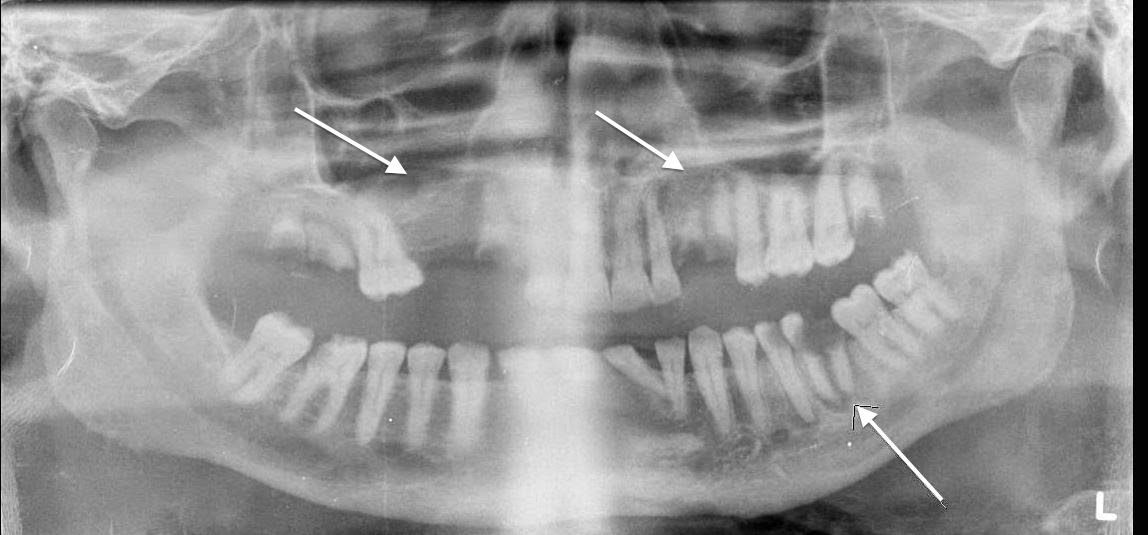
53 year old gentleman had chest X ray for as part of some routine workup, which is shown below


The chest X ray showed a calcified well circumscribed mass of about 3 cm in size, as pointed by the arrow.

Differential Diagnosis include

Malignancy which could be primary lung or metastatic
Tuberculosis
Fungal infections

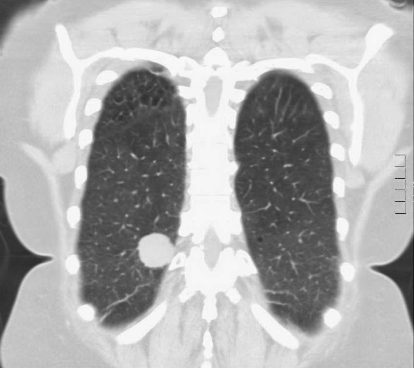
CT chest was ordered and shown below


CT scan confirmed the findings and showed well circumscribed, calcified mass with clear borders and Golf Ball like appearance. 

Biopsy showed spindle shaped cells and yeast forms, confirming the diagnosis of histoplasma. The well circumscribed golf ball like mass is known as histoplasmoma. 

Since this pt was asymptomatic, No treatments interventions were performed


Sunday, March 2, 2014

Opacification and Sclerosis of Sphenoid Sinus

Image below shoes thickening and sclerosis of sphenoid sinus wall (as shown by arrow). This shows long standing inflammatory process. Differential include chronic bacterial or fungal sinusitis